25 Stunning Photographs of the Fetus Developing by Lennart Nilsson - Embryo Photos
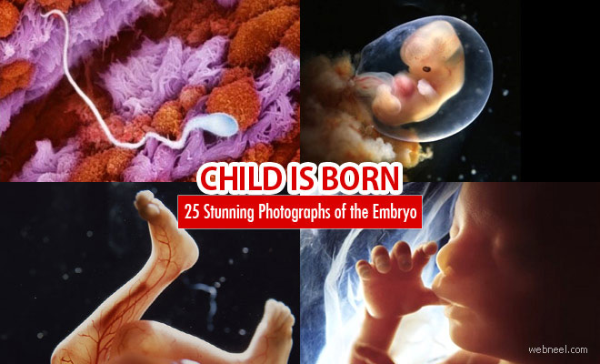
Embryo / Fetus Photos: Seeing a newborn makes every mother smile, but the joy can be even greater when parents see images of their babies as embryos. Mothers want to capture every moment of their child's life, so from the first heartbeat or visible life form on an ultrasound scan, they often want to preserve these memories forever. Embryo photography is sought after by many couples who want to share these special moments with their child when they grow up. In this post, we have included 25 beautiful embryo photos for your inspiration. Embryo photography shows the week-by-week development of the fetus from a tiny living cell to a full-sized baby in the mother's womb.
Embryo
Egg cell
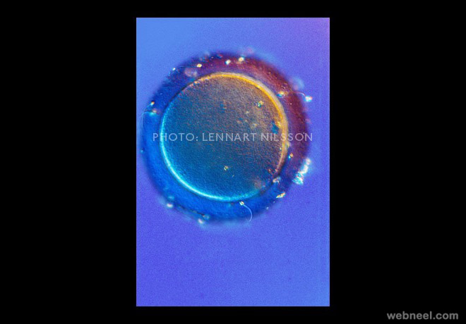
Egg sperm
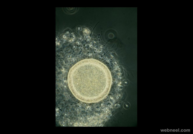
Millions of sperm entering the vagina and swim towards the opening of the cervix.
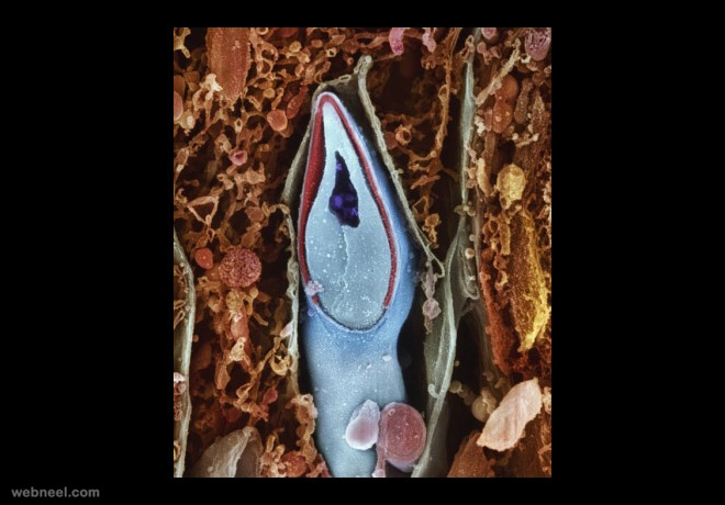
Sperm fallopian tube
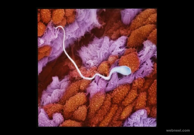
The winning sperm
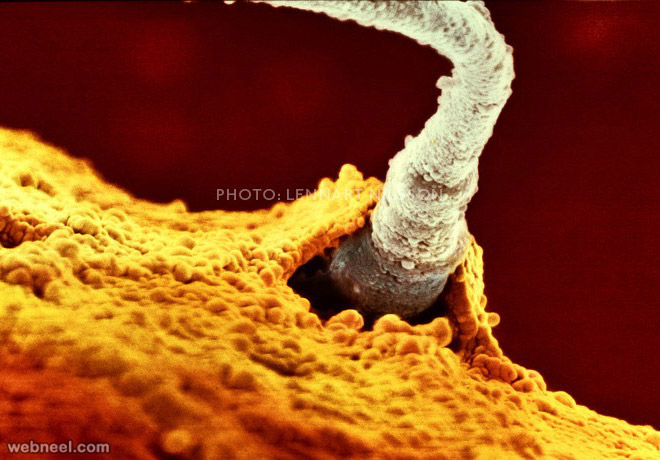
Embryo 6days : 5-6 days, the clump has developed into a blastocyst, containing many more cells, and has entered the womb
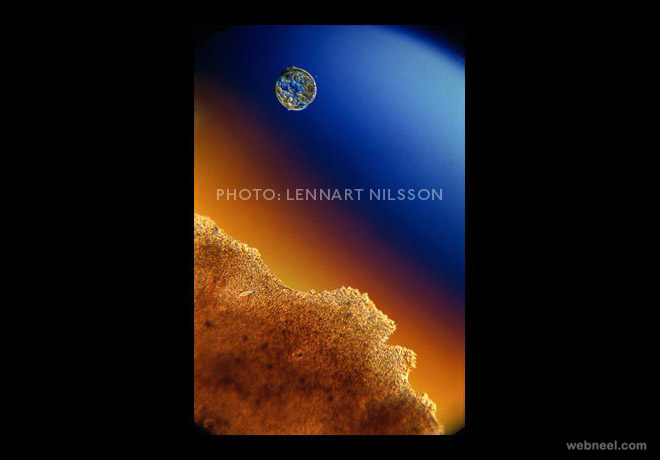
Embryo 8days: The rapidly-growing embryo is well protected in the foetal sac
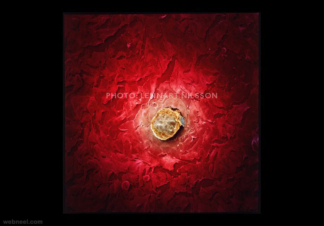
Embryo 4weeks
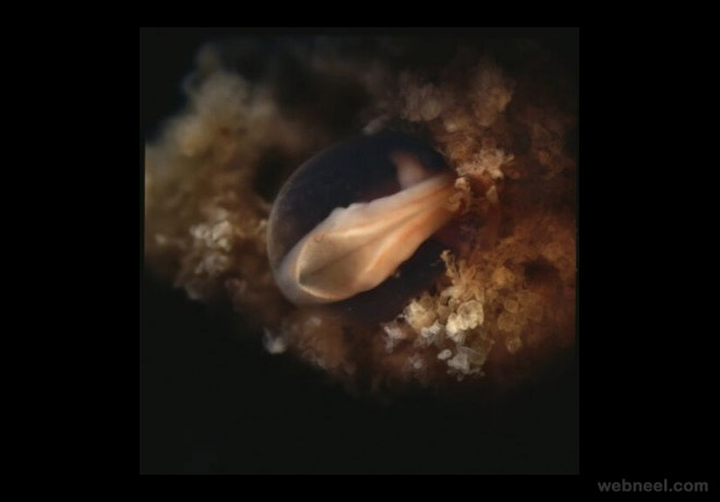
Embryo 5weeks : The embryo is approximately 9mm long. A face develops, with openings for the mouth, the nostrils and eyes
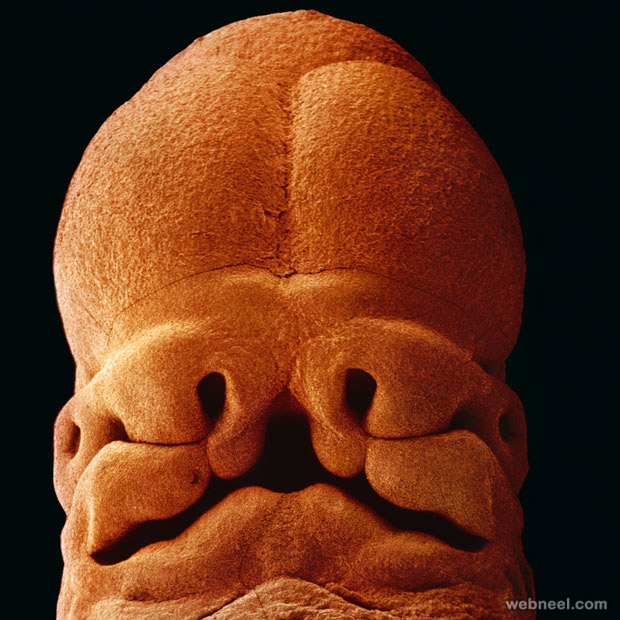
Fetus 6weeks
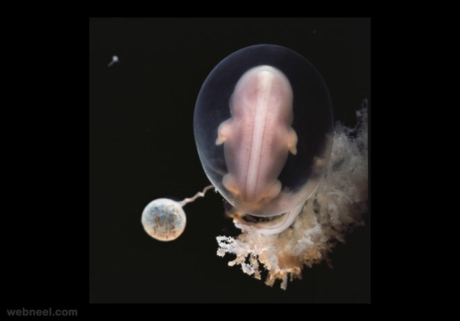
Fetus 7weeks
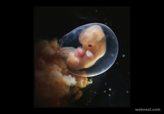
Fetus 10weeks : The eyelids are semi-shut. They will close completely in a few days
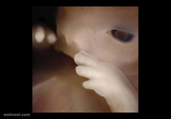
Spaceman 13weeks
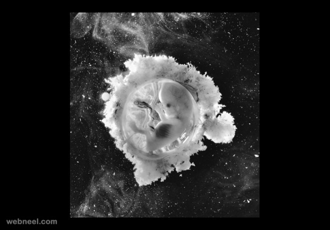
Fetus 16weeks: The foetus uses its hands to explore its own body and its surroundings
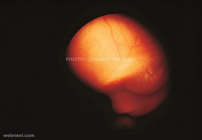
The foetus can now grab and pull the long umbilical cord. The skeleton consists mainly of flexible cartridge. A network of blood vessels is visible through the thin skin
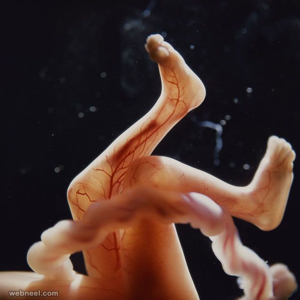
18 weeks: Approximately 14cm. The foetus can now perceive sounds from the outside world
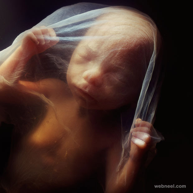
Fetus 19weeks

Fetus 20weeks

Embryo 20weeks

20 weeks. Approximately 20cm. Woolly hair, known as lanugo, covers the entire head

Foot of an embryo
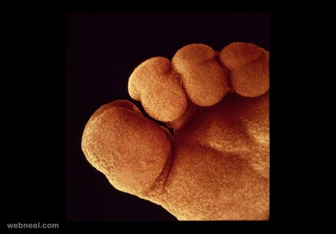
Sperm entering
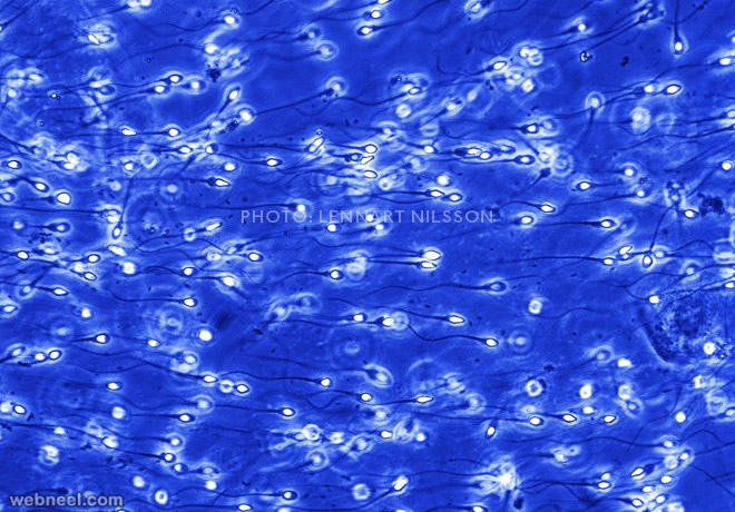
Fallopian tube
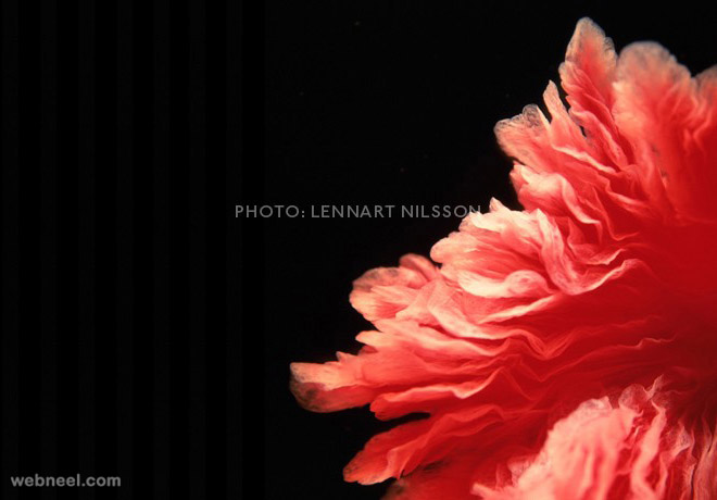
Embryo heart cells
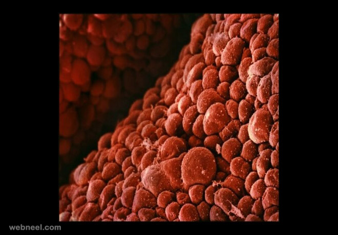
Embryo laguno hair
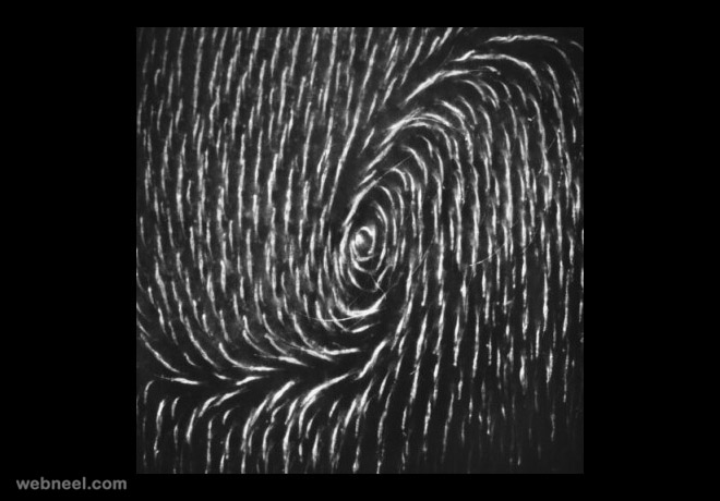
Embryo cervix

Know more about Lennart Nilsson